CELL PAINTING
ASSAY SERVICES


STEP 1
Culture cells and treat with compound of interest.

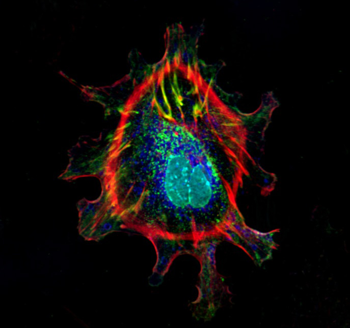
STEP 2
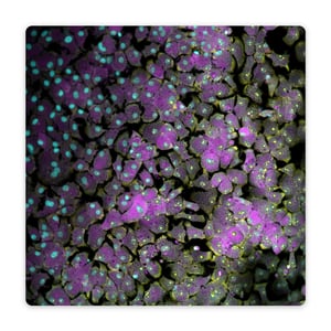
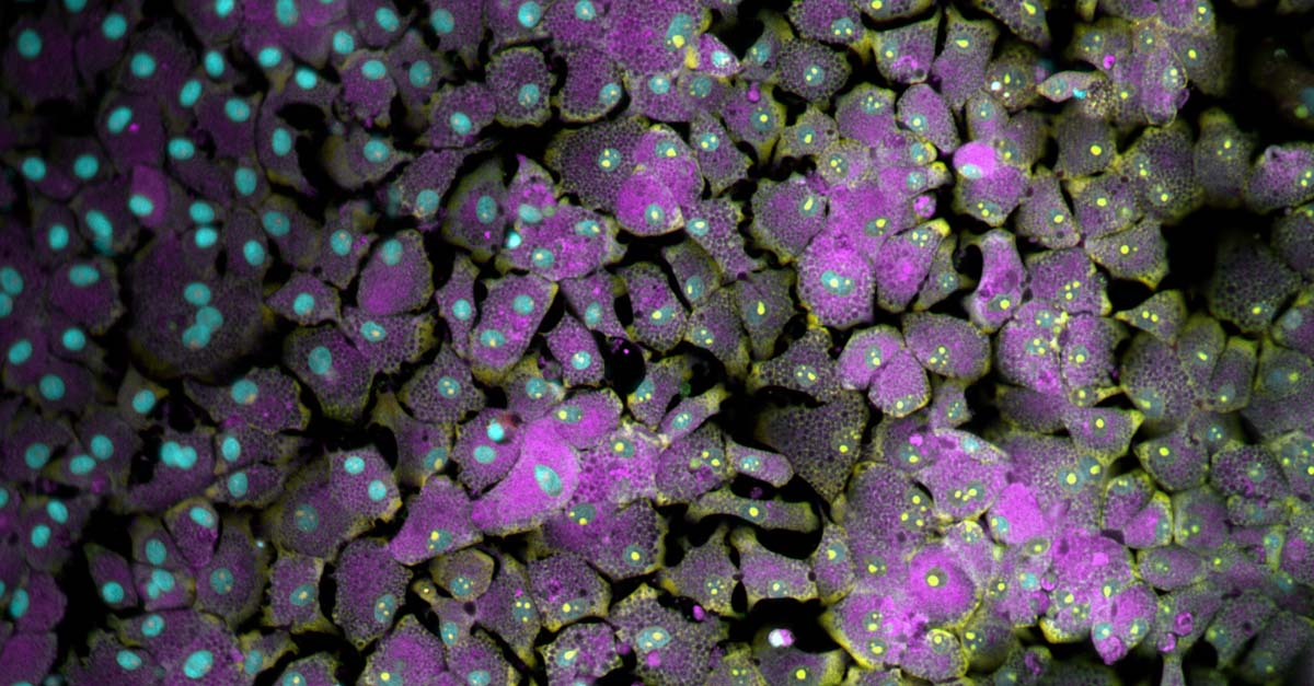
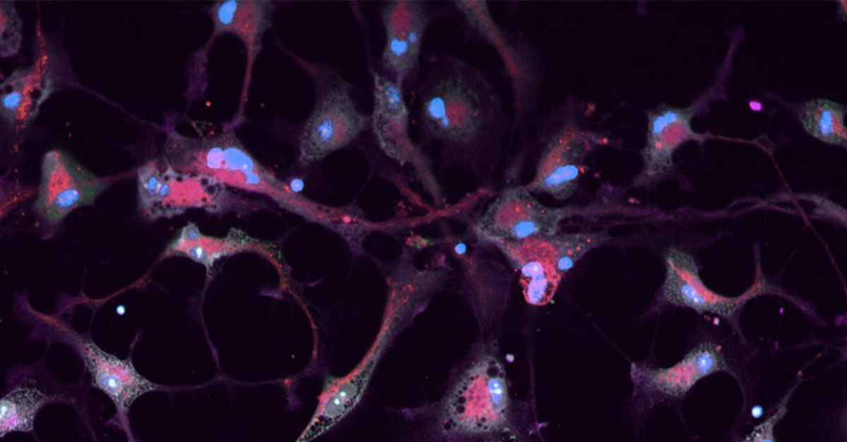
Stain cells with a palette of fluorescent dyes that target different cellular structures.

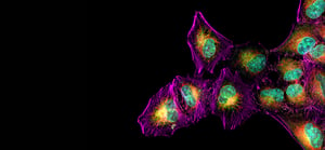
STEP 3
Capture high-resolution images of stained cells using fluorescence microscopy.

STEP 4
Analyze data to identify phenotypic changes and compare against a reference library.
STEP 1
Culture cells and treat with compound of interest.
STEP 2
Stain cells with a palette of fluorescent dyes that target different cellular structures.
STEP 3
Capture high-resolution images of stained cells using fluorescence microscopy.
STEP 4
Analyze data to identify phenotypic changes and compare against a reference library.
Cell Painting in Preclinical Safety and Toxicology: Literally Taking a Closer Look
Cell Painting offers immense potential for the future of drug discovery. Learn more about this powerful, imaging-based technique and how it can enhance preclinical safety and toxicology assessments of therapeutic candidates.