In the field of drug development, understanding the intricate mechanisms by which a drug interacts with cells is crucial. One powerful technique that has gained significant traction in recent years is cell painting. This innovative approach involves the comprehensive profiling of cellular responses to compounds or drug candidates, enabling scientists to unravel elusive mechanisms of action (MOAs) and expedite the drug-discovery process. In this post, we discuss how cell painting is revolutionizing the way drugs are developed and evaluated.
TABLE OF CONTENTS
Using Cell Painting to Determine Mechanism of Action
The Advantages of Cell Painting in Drug Development
The Basics of Cell Painting
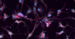
Cell painting is a high-content screening technique that employs fluorescent dyes to label various cellular components, enabling researchers to visualize and quantify cellular changes through high-resolution imaging. This technique relies on the principle that drugs affect different cellular structures and processes in a way that results in distinct, phenotypic changes.
By using a combination of fluorescent dyes that target and “paint” specific subcellular compartments or structures, such as the nucleus, cytoplasm, and mitochondria, scientists generate detailed, systemic views of how various treatment paradigms affect the overall phenotypic features of cells. The differences in cellular phenotypes highlight the alterations induced by specific treatments.
Fluorescent signals emitted by the dyes are captured using advanced, high-content microscopy techniques, and image analysis allows for the quantification of thousands of cellular features. Unique combinations of phenotypic features for each treatment paradigm allow scientists to assign phenotypic “fingerprints” that represent each experimental condition.
Using Cell Painting to Determine Mechanism of Action
One of the primary challenges in drug development is deciphering how a drug exerts its therapeutic effects on cells. Understanding the MOA is crucial for optimizing drug efficacy and minimizing adverse effects. Cell painting offers an effective approach to unraveling MOA by examining cellular-response patterns induced by different compounds.
Using machine learning algorithms and computational analysis, large-scale datasets generated by cell painting can be analyzed to identify distinct phenotypic profiles - or fingerprints - associated with specific MOAs. By comparing these phenotypic profiles to a database of known MOAs, scientists can infer the mechanism by which the novel drug exerts its effects, providing valuable insights for subsequent stages of drug development. This approach is particularly valuable for compounds with unknown or poorly characterized MOAs, as it provides a systematic and unbiased evaluation of cellular response. Moreover, cell painting can uncover subtle changes that may be overlooked or undetectable by traditional methods, offering a more comprehensive understanding of a drug's effects.
The Advantages of Cell Painting in Drug Development
- Comprehensive Profiling: Cell painting allows for the simultaneous evaluation of multiple cellular features, providing a holistic view of drug-induced changes. This comprehensive profiling enables researchers to identify both intended and unintended effects of a drug, aiding in the optimization of therapeutic efficacy.
- Early-stage Screening: Cell painting can be employed in the early stages of drug development, allowing researchers to quickly assess the potential MOA of a compound. This early insight can help prioritize lead compounds, saving valuable time and resources in the drug-discovery process.
- Translational Relevance: Cell painting can bridge the gap between in vitro and in vivo models by capturing complex, cellular responses, using physiologically relevant, human cell types (e.g., iPSC-derived or primary cells). This translational relevance enhances the predictability and applicability of the drug candidate in the clinic, ultimately improving the success rate in clinical trials.
- Reproducibility & Scalability: One of the major advantages of cell painting is its scalability. With the advent of high-throughput technologies and automated microscopy systems, researchers can analyze thousands of compounds simultaneously, generating massive datasets. These datasets can be subjected to advanced, image-analysis algorithms and machine-learning techniques, enabling the identification of novel MOAs and the discovery of potential therapeutic targets. Furthermore, cell painting can help identify off-target effects and potential toxicity issues at an early stage of drug development.
Conclusion
In the realm of drug development, cell painting has emerged as a valuable tool for deciphering the MOA of novel compounds. By combining advanced microscopy and computational analysis, researchers can generate comprehensive profiles of cellular responses, unraveling the intricate mechanisms underlying drug action. The ability to identify and prioritize lead compounds early in the drug-discovery process, while also gaining insights into potential adverse effects, is transformative for the field. As cell painting continues to evolve, it holds great promise for accelerating drug development and improving patient outcomes.
References
- Bray, M., Carpenter, A., Kaini, R., et al. (2017). Advanced assay development guidelines for image-based high content screening and analysis. In: Assay Guidance Manual. Bethesda (MD): Eli Lilly & Company and the National Center for Advancing Translational Sciences.
- Perlman, Z. E., Slack, M. D., Feng, Y., et al. (2004). Multidimensional drug profiling by automated microscopy. Science, 306(5699), 1194-1198.
- Bray, M. A., Gustafsdottir, S. M., Ljosa, V., et al. (2017). A dataset of images and morphological profiles of 30,000 small-molecule treatments using the Cell Painting assay. GigaScience, 6(12), 1-5.
- Caicedo, J. C., Singh, S., Carpenter, A. E. (2016). Applications in image-based profiling of perturbations. Current Opinion in Biotechnology, 39, 134-142.