Cell Painting offers immense potential for the future of drug discovery as a powerful, imaging-based technique commonly used for preclinical safety and toxicology assessments of therapeutic candidates.
TABLE OF CONTENTS
Cell Painting in Preclinical Safety and Toxicology
1. Assessment of Cellular Morphology
2. Unraveling Intracellular Signaling Pathways
3. Quantifying Cellular Responses
A Promising Future for Drug Discovery
WHAT IS CELL PAINTING?
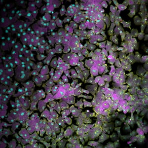
Cell painting is an imaging-based technique that allows scientists to capture the intricate details of cellular structures and functions, including general measurements of size, shape, and location, to nuanced information, such as texture and intensity, by analyzing images of cells stained with a palette of dyes that target various components of cells (e.g., nuclei, cytoskeleton, individual organelles). Rich datasets with thousands of feature measurements can be extracted from images to analyze the effects of treatments on the basic functions and structures of the cells.
Unique combinations of these feature measurements that result from a particular treatment are akin to a “fingerprint” of that cellular state. Differential phenotypic profiles are illustrated in the figure below, in which A549 cells were treated with bafilomycin or cytochalasin D and stained with a five-channel palette. Similar or matching fingerprints that result from different treatments may be indicative of a similar mechanism of action. The bigger your database of cellular fingerprints, the better the chance of matching fingerprints, and the closer you are to understanding a compound’s effects.
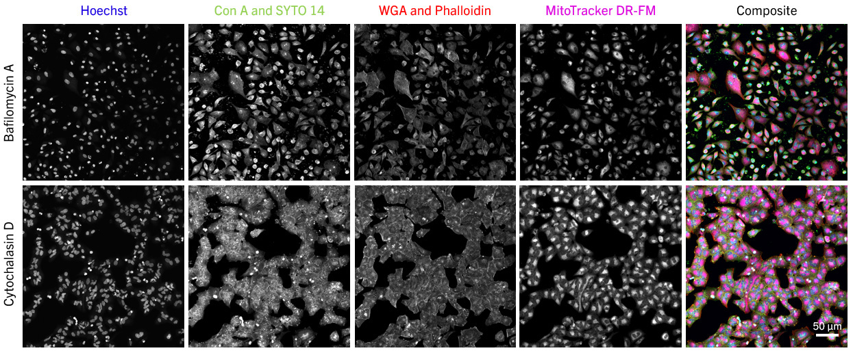
A549 cells show different phenotypic profiles when treated with bafilomycin A vs cytochalasin D and stained with the standard cell-painting palette.
Cell Painting is often performed on routine cell types (think A549 or U2-OS), but it can also be performed on more clinically relevant models, such as iPS-derived cell types. For example, for cardiotoxicity studies, this technique can be performed on cardiomyocytes. In liver toxicity studies, it can be run with hepatocytes.
CELL PAINTING IN PRECLINICAL SAFETY AND TOXICOLOGY
In the quest for safer and more effective drugs, cell painting plays a crucial role in high-throughput toxicity screening. By exposing cells to a library of compounds and analyzing their effects, scientists can identify potential toxic responses or discover off-target effects of existing drugs. Screening and comparing drug candidates to libraries of known toxicants/compounds provides valuable insights into their potential, toxic effects which can be broadly categorized as follows:
1. ASSESSMENT OF CELLULAR MORPHOLOGY
Cell painting provides a visual fingerprint of cellular changes induced by therapeutic candidates. This involves characterizing alterations in cellular and subcellular shape, size, and structure that result from treatment.
2. UNRAVELING INTRACELLULAR SIGNALING PATHWAYS
Therapeutic candidates can have a range of intended and unintended consequences that can wreak havoc on the signaling pathways within cells. With cell painting, we track the effects of candidates on specific targets and compare them to the effects of compounds with known mechanisms of action. This information can tell us to which pathways are being disrupted.
3. QUANTIFYING CELLULAR RESPONSES
With cell painting, we quantify the effects of therapeutic candidates at a cellular level. By analyzing the intensity and distribution of cellular stains, we can measure changes in key parameters like cell viability, proliferation, cellular morphology, or metabolic activity. This quantitative data helps us understand dose-response relationships and identify mechanisms of action and off-target effects.
A PROMISING FUTURE FOR DRUG DISCOVERY
As we unlock the potential of cell painting in preclinical safety and toxicology, we realize its game-changing impact. By providing a visual and quantitative understanding of cellular responses to therapeutic candidates, cell painting helps us better comprehend their mechanisms of action and safety profiles. This not only accelerates the process of bringing life-saving treatments into the clinic, but also instills confidence in their safety.
REFERENCES:
- Bray, M.-A. et al. (2017). Cell Painting, a high-content image-based assay for morphological profiling using multiplexed fluorescent dyes. Nature Protocols, 11(9), 1757-1774.
- Cosgrove, B. D. et al. (2017). A high-throughput screening assay for assessing the viability and cytotoxicity of 3D-printed materials. ACS Biomaterials Science & Engineering, 3(8), 1472-1481.