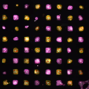
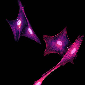
A client requested a custom, neuronal model to evaluate their therapeutic agent’s effects on tau expression and phosphorylation using iPSC-derived glutamatergic neurons from two different donor backgrounds.
Goals:
- Establish an assay to measure tau expression and phosphorylation in healthy and MAPT P301S human, iPSC-derived glutamatergic neurons.
- Evaluate the effects of client’s therapeutic agent on various measures of tau in each culture model.
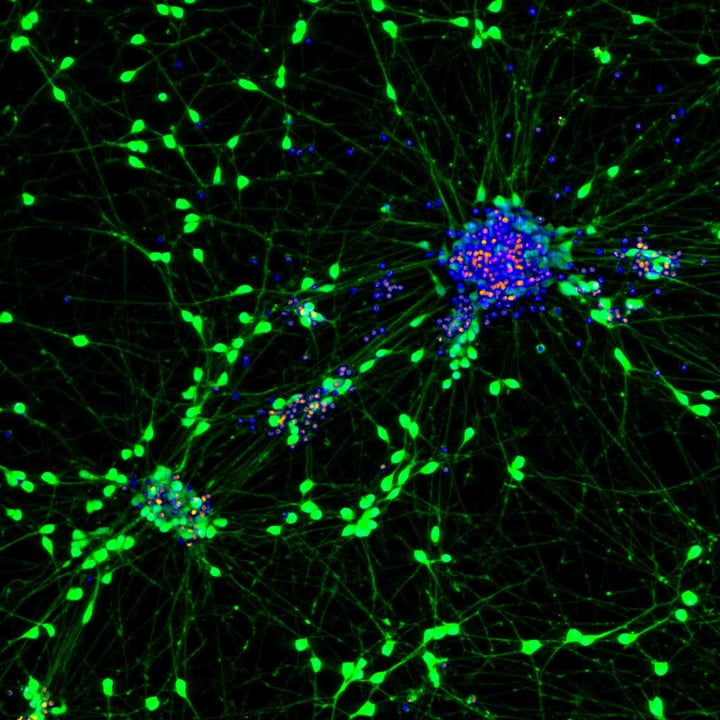
EXPERIMENTAL DESIGN
Cell Model
iPSC-glutamatergic neurons
- WT/WT
- MAPT P301S
Palette 1
Hoechst (nuclei)
Tuj1
Total Tau
Phospho-tau Ser202/Thr205 (AT8)
Palette 2
Hoechst
Tuj1
Total Tau
Phospho-tau Thr217 (6G7C8)
Treatments and Timelines
- Culture cells in 384-well, imaging microplates, following custom culture protocol developed at PhenoVista until DIV12.
- On DIV12, treat with test articles
- Continue with half-media changes every other day for two weeks, readministering treatments with each change.
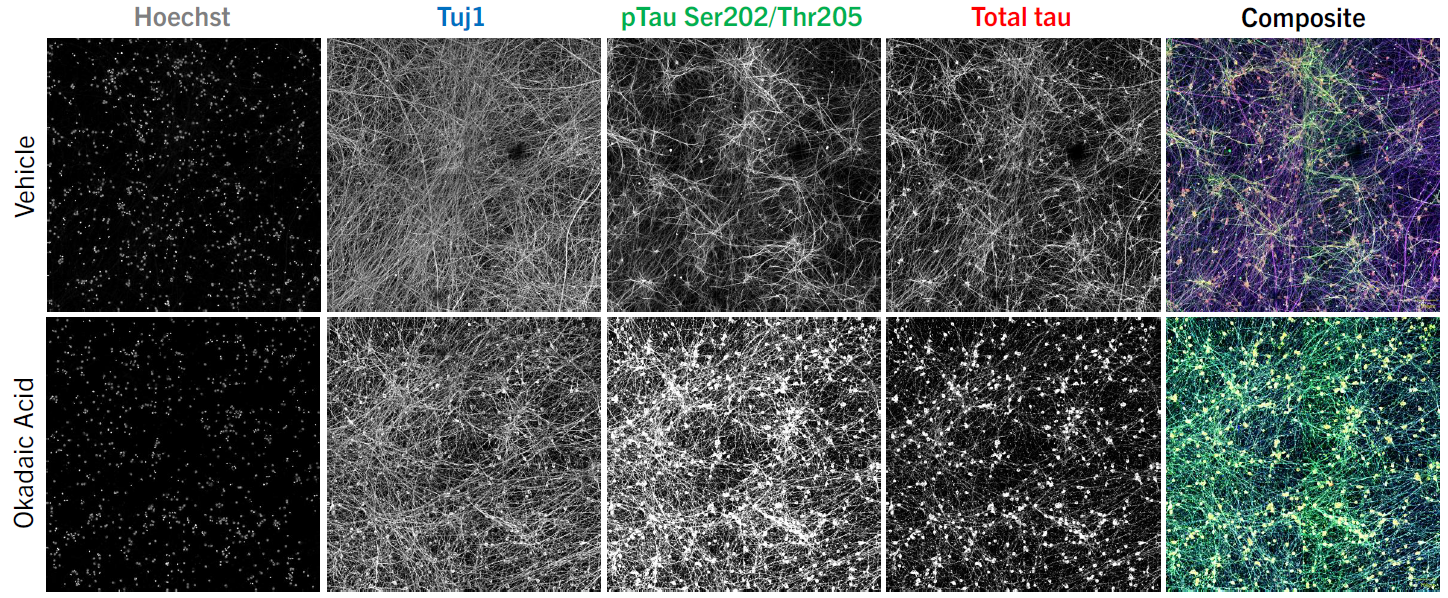
- On DIV26, treat appropriate control wells with okadaic acid (inhibitor of serine/threonine protein phosphatases, causing tau hyperphosphorylation) 3hrs prior to fixation.
- Fix and stain cultures for scanning and analysis.
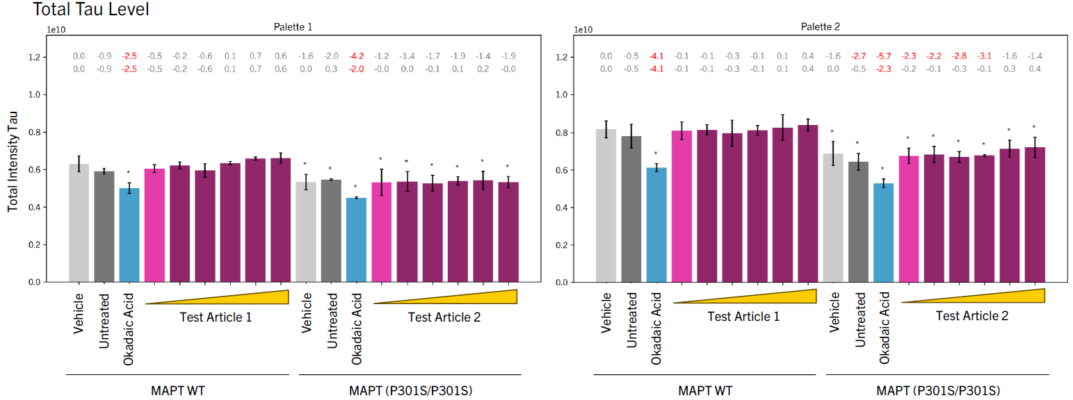
REPRESENTATIVE IMAGES & QUANTITATIVE DATA


RELATED RESOURCES
CONTACT US
Let's discuss how we can help with your next study.
DOWNLOAD THE CASE STUDY
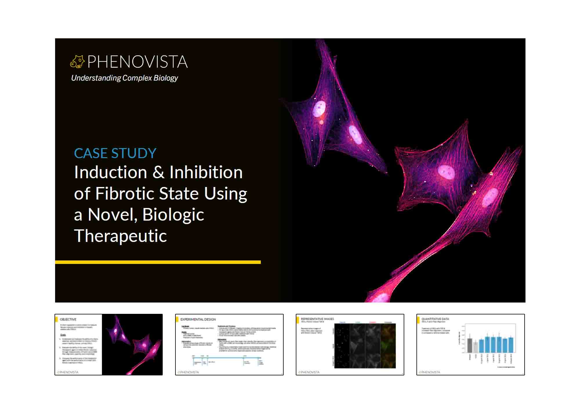
Induction & Inhibition of Fibrotic State Using a Novel, Biologic Therapeutic
Overview
Review an example of a study designed to measure fibrosis induction and inhibition in hepatic stellate cells (HSCs).
Highlights
- Overview of study objective and goals
- Experimental design, including timelines and deliverables
- Representative images and graphs
- Study results
![]()