
READY-2-GO
LUNG FIBROSIS
ASSAY SERVICE
Measure fibrotic induction and inhibition in human, primary lung fibroblasts.
KEY FEATURES
![]()
Uses human, primary cells in an in vitro model of lung fibrosis
![]()
Readouts that characterize intracellular and extracellular fibrosis indicators
![]()
Only 6-8 weeks from assay to report
![]()
Option to bundle R2G Services or transition to Custom Services
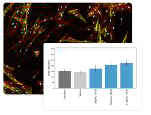
ASSESS YOUR THERAPEUTICS' EFFECT ON FIBROTIC INDUCTION
Primary, human, normal lung fibroblasts are treated with your test articles prior to fibrotic induction, and images are acquired and analyzed at various time points.
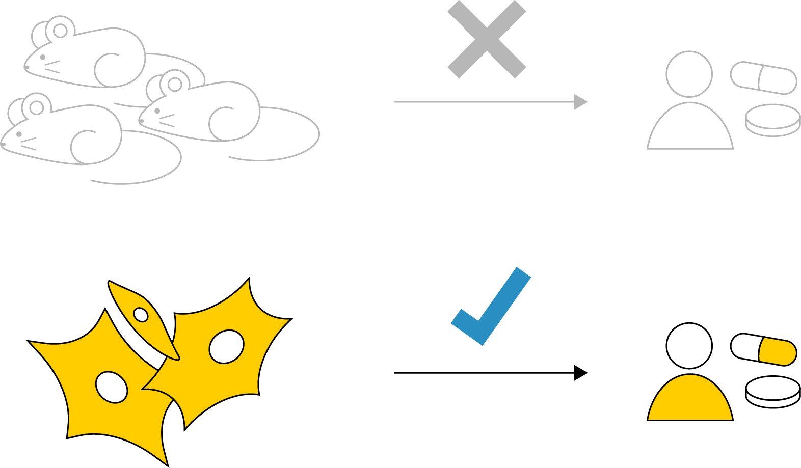
AVOID SPECIES DIFFERENCES WITH PRIMARY, HUMAN, LUNG FIBROBLASTS
The use of animal models or immortalized cell lines limits clinical translatability. We use primary, human, lung fibroblasts to help bridge the gap between the bench and clinic.
RELATED READY-2-GO SERVICES
Browse related Ready-2-Go assay offerings. For custom assays, check out our Custom Services.
![]()
R2G CELL HEALTH
Multiplexed readouts of proliferation, viability, and apoptosis.
Learn more
![]()
R2G MITOCHONDRIAL HEALTH
Screen for changes in mitochondrial membrane potential.
Learn more
![]()
R2G LIVER FIBROSIS
Fibrotic induction and inhibition are measured in stellate cells.
Learn more