This blog entry is contributed by Carla Neiser from Sapient Bioanalytics.
Table of Contents
HCI for Discovering and Assessing Phenotypes
Multi-omics for Differentiating Mechanisms within Phenotypes
Practical Applications of Integrated HCI-Omics Approaches
Conclusion: Mapping Biological Complexity to Inform R&D Decisions
Introduction
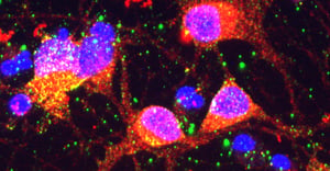
High-content imaging (HCI) techniques enable high-dimensional cell analysis at scale, extracting hundreds to thousands of quantitative features per cell across millions of cells at a time. Combining readouts of cellular morphology, protein localization, organelle structure, cell-population metrics, and expression levels, HCI delivers a comprehensive view of how cells respond to perturbations from genetic mutations to treatments with compounds. Unlike targeted assays, which focus on a single, known endpoint, HCI allows investigators to broadly observe cellular changes and identify unexpected or novel biology, making it ideal for phenotype-driven discovery.
While HCI is a powerful tool to answer “what” cellular structures and behaviors visibly change after a perturbation – and to cluster those conditions based on phenotypic similarity – it can be made even more powerful with the addition of orthogonal, multi-omics data that provide unprecedented insight into the “why,” such as mapping pathway‑level mechanisms and molecular drivers behind the HCI-observed signatures. Genetic mutations or various stimuli may produce similar morphological effects in cells, making them look nearly identical under HCI, but each may act through distinct molecular pathways. Multi‑omics provides the mechanistic resolution needed to deconvolute these ambiguities.
HCI for Discovering and Assessing Phenotypes
HCI enables measurement of multivariate cell states at single‑cell resolution and scale, enabling unbiased profiling of drug effects, genetic perturbations, toxicity, and cell health. These rich phenotypic signatures map functional outcomes in ways that traditional, single‑parameter assays cannot, especially for complex diseases in which mechanisms are distributed across multiple pathways, rather than restricted to a single target. Large-scale studies have validated that HCI‑derived morphological profiles can predict compound bioactivity and mechanism classes, establishing it as a robust technique for discovery.
Still, phenotypic similarity does not guarantee mechanistic equivalence. Compounds that produce nearly identical imaging-derived phenotypes can do so for very different reasons; for example, one may activate a DNA‑damage checkpoint, while another may rewire mitochondrial metabolism. While HCI shows the outcome of upstream molecular events, additional multi-omics data are needed to identify the processes that produced that outcome, so that the relevant pathways can be further acted upon.
Multi-omics for Differentiating Mechanisms within Phenotypes
Integrated analyses combining HCI and multi-omics are increasingly recognized as essential for mechanistic discovery and deconvolution. Modern mass spectrometry-based workflows can directly quantify thousands of proteins, metabolites, and lipids across hundreds of pathways and biological processes in a single sample, enabling more precise mechanism‑of‑action mapping and capture of disease-relevant processes that underlie observed phenotypic changes.
Proteomics, for example, quantifies changes in protein abundance and post‑translational modifications (PTMs), revealing which signaling networks are engaged, activated, or disrupted. Studies in neurodegeneration and cancer reveal that proteomic signatures often align with and also refine phenotypes detected by HCI, identifying the specific pathways that drive morphological change. Similarly, metabolomics and lipidomics capture shifts in metabolic flux, energy balance, and lipid signaling that frequently underlie organelle morphology, mitochondrial health, and cell‑state transitions observable by HCI.
By overlaying molecular changes onto HCI-derived phenotypic clusters, researchers can determine not only how conditions group together, but why they group that way - whether through shared, upstream targets, convergent stress responses, compensatory signaling, or entirely distinct mechanisms that happen to produce similar phenotypic readouts.
Practical Applications of Integrated HCI-Omics Approaches
Across therapeutic areas, the combination of HCI and multi-omics data can be used to capture rich, multiparametric phenotypes and their underlying molecular drivers, teasing apart look‑alike phenotypes to reveal which pathways are actually driving the observed responses.
CNS Diseases
In CNS research, HCI enables precise quantification of synaptic density, neurite outgrowth, mitochondrial integrity, and other structural or functional hallmarks of neuronal health. These phenotypes are powerful, but imaging alone cannot reveal the upstream drivers of those changes. By adding comprehensive proteomic measures across 10,000+ critical proteins, including through cytokine profiling, researchers can detect shifts in synaptic scaffolding proteins, neuroinflammatory mediators, and other protein-mediated functional biology that shape neuronal responses. When combined, HCI and multi‑omics provide a unified framework for understanding why CNS‑related phenotypes improve, worsen, or diverge, even when neuronal morphology or network structure appear similar by imaging alone.
Oncology
In oncology, HCI captures a wide array of tumor-cell behaviors, including proliferation rates, cell‑cycle transitions, apoptotic signatures, and organelle stress, offering a granular view of how cancer cells respond to treatment. Multi‑omic approaches like proteomics and metabolomics enable mechanistic differentiation within a given phenotype by revealing whether such behaviors are driven by DNA‑damage signaling, metabolic collapse, rewiring of key metabolic pathways, immune‑modulatory activity, or other upstream processes. Mechanistic context is essential in cancer biology, where phenotypically similar imaging signatures can mask profoundly different biological drivers – each with unique therapeutic implications.
Fibrosis
For fibrosis models, HCI provides comprehensive measurement of extracellular matrix (ECM) deposition, collagen maturation, myofibroblast activation, and other structural outcomes that define fibrotic progression. Proteomics adds upstream clarity by identifying alterations in pathway signaling, ECM‑interactome dynamics, and fibroblast-specific protein expression, while metabolomics further reveals shifts in energy utilization and lipid‑mediated signaling that influence fibroblast activation and ECM production. Together, HCI and multi‑omics reveal whether an antifibrotic compound acts upstream of canonical signaling, downstream on matrix assembly, or through entirely alternative metabolic or molecular mechanisms, offering a far more complete picture of therapeutic impact.
Conclusion: Mapping Biological Complexity to Inform R&D Decisions
By combining HCI and multi-omics within well‑tuned, human‑relevant models, researchers can map a faster route from phenotypic observation to mechanistic insight and biomarker discovery, building greater confidence in findings along the way. HCI‑detected morphological signatures in disease models can be tied to altered protein networks, and HCI‑observed clusters of drug responses can be annotated with pathway‑level insight. Novel phenotypes identified through unbiased imaging can be mechanistically explained through targeted or global -omics analysis. This integration builds upon HCI as a discovery engine, closing the loop between phenotype and proof of mechanism to increase relevance and translatability while reducing the risk of false conclusions from single-omic measures.