READY-2-GO
MICROGLIA PHAGOCYTOSIS
ASSAY SERVICE
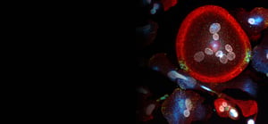
Measure the uptake of bacterial bioparticles by human, iPSC-derived microglia.
KEY FEATURES
![]()
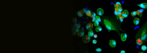
Uses wild-type or TREM2-mutant, human, iPSC-derived microglia

Quantitative, live-cell and endpoint measurements of phagocytosis
![]()
Only 4-6 weeks from assay to report
![]()
Option to bundle R2G Services or transition to Custom Services
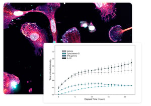
ASSESS YOUR THERAPEUTICS' EFFECT ON PHAGOCYTOSIS ACTIVATION
iPSC-microglia are treated with reference compounds that promote or inhibit activation of phagocytosis alongside your test articles. Fluorescence of bacterial bioparticles is measured every hour for 24 hours.
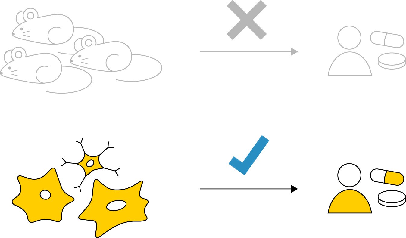
AVOID SPECIES DIFFERENCES WITH HUMAN, iPSC-DERIVED MICROGLIA
The use of animal models or immortalized cell lines limits clinical translatability. Choose from wild-type, TREM2-homozygous, or TREM2-heterozygous, human, iPSC-derived microglia for a scalable and clinically relevant model.
RELATED READY-2-GO SERVICES
Browse related Ready-2-Go assay offerings. For custom assays, check out our Custom Services.
![]()
R2G NEUROTOXICITY
Multiplexed readouts of viability and apoptosis.
Learn more
![]()
R2G NEURONAL MITOCHONDRIAL HEALTH
Multiplexed readouts of mitochondrial health.
Learn more
![]()
R2G NEURITE OUTGROWTH & NEURITE NETWORK DYNAMICS
Quantification of neurite outgrowth and changes in established neurite networks using human, iPSC-derived neurons.
Learn more