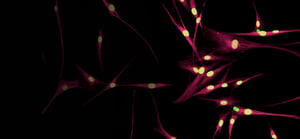
HIGH-CONTENT IMAGING
High-content imaging is a powerful technique for measuring and monitoring phenotypic changes in cells in early drug discovery.
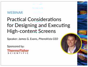
Practical Considerations for Designing and Executing High-content Screens
Learn about the basic principles and process of high-content screening with tips on how to design a successful assay.
Speaker: James G. Evans, PhenoVista CEO
WHAT IS HIGH-CONTENT IMAGING USED FOR?
HCI is used across all areas of biology, from cell and molecular biology to drug screening, and its primary objective is to extract multi-dimensional data from single assay runs through three main steps - image acquisition, image processing, and image analysis. With one in vitro experiment, HCI affords quantitative, phenotypic datasets to evaluate the performance of diverse therapeutic modalities at the single-cell and subcellular levels and provides researchers an understanding of underlying biological heterogeneity across multiple readouts simultaneously, such as pathway activation, cellular localization/translocation, and morphology. The detailed resolution of such readouts allows researchers to explore complex biological questions.
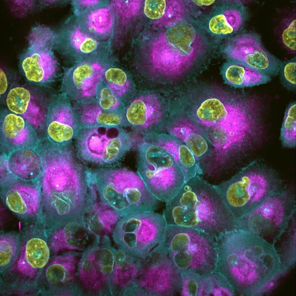

APPLICATIONS OF HIGH-CONTENT IMAGING
HCI is not limited to simple cell models; it can be applied to sophisticated in vitro models, including co-cultures of cell types in 2D and 3D and microfluidic devices. HCI can be performed in live or fixed cells and adapted to medium- and high-throughput formats. The flexibility and broadness of this technique allows researchers and drug developers to customize assays to fit their specific needs and answer questions pertaining, but not limited, to cytotoxicity, target identification, target validation, therapeutic efficacy, mode-of-action determination, and therapeutic delivery.
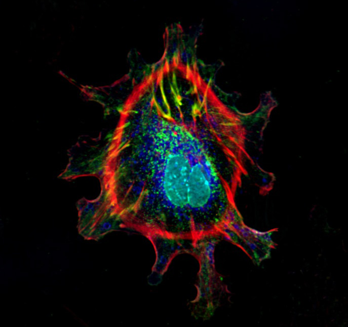
ASSAY SERVICE OFFERINGS
Our expertise lies in the use of physiologically relevant cell models and high-content imaging to get you actionable insights into the effects of your therapeutic agents in various areas of disease research.
![]()
CUSTOM ASSAYS
Custom assays to answer more specific, complex biological questions.
Learn more
![]()
READY-2-GO
Defined, fixed offerings across a range of disease and therapeutic areas.
Learn more
![]()
CELL PAINTING
Handle a large number of compounds and compare against known drugs.
Learn more
![]()
IMAGING & ANALYSIS
You send us plates with cells that have been fixed & stained, we'll send you data.
Learn more